
Image

Our sponge collection was started in 1968 under Dr David Erwin as part of an inventory of Northern Ireland's marine animals. It was expanded as part of an initiative by the Marine Conservation Society to create an identification guide for divers to record which species they were seeing underwater.
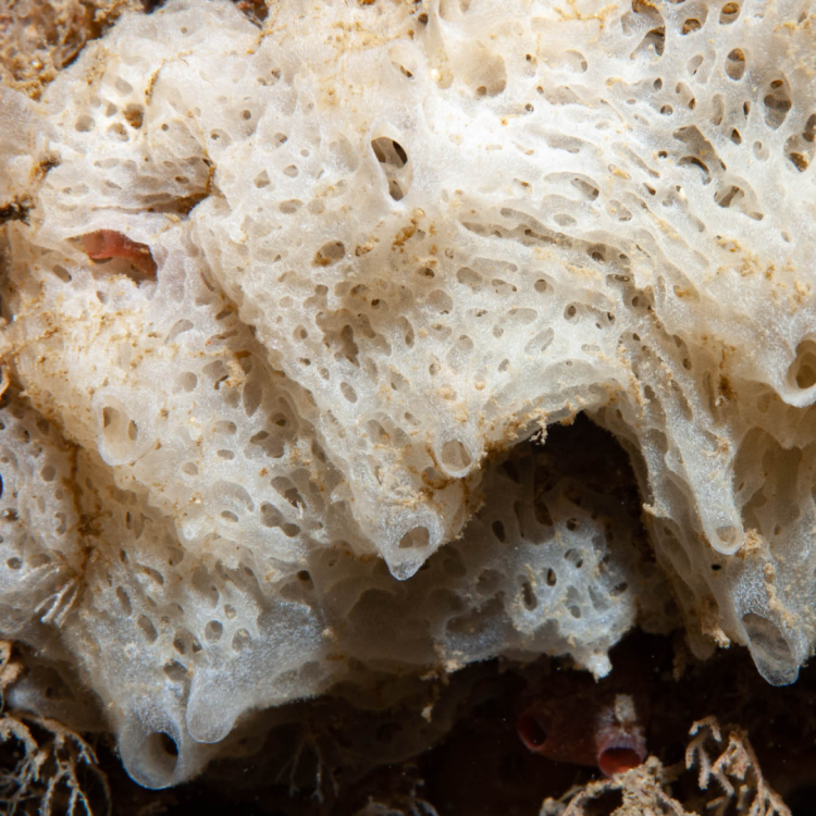
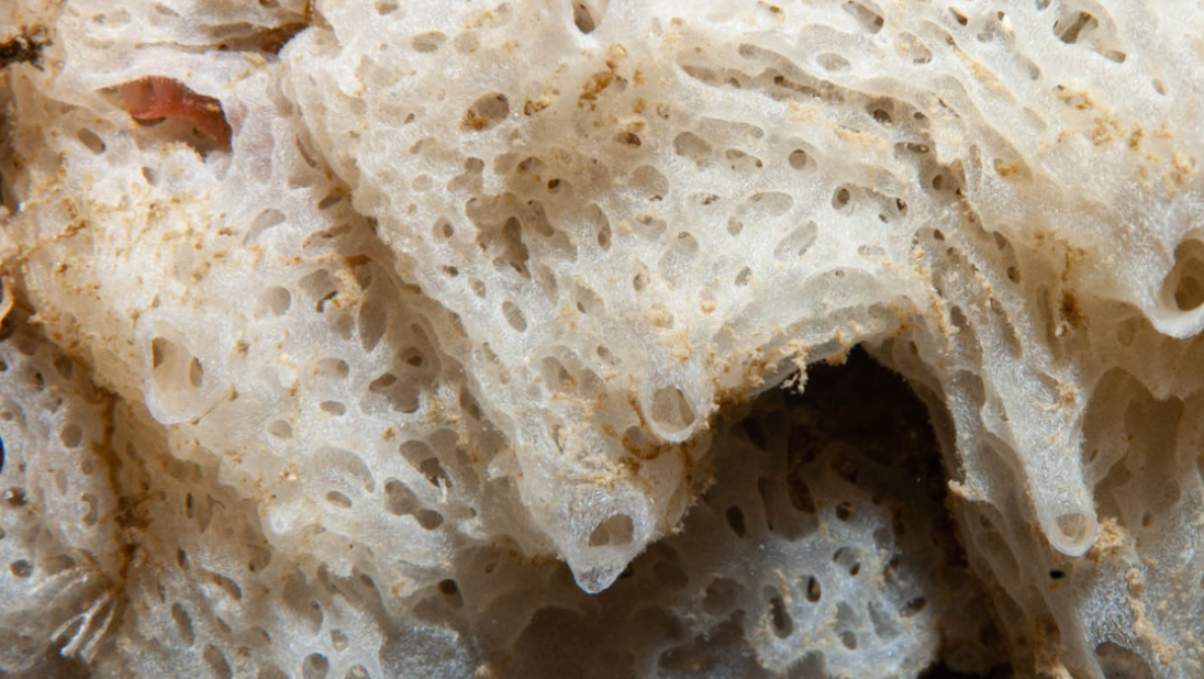
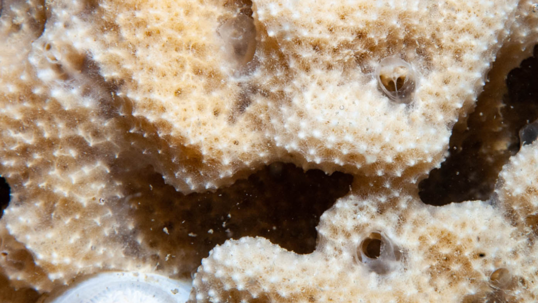
Sponges are a diverse group of animals, with about 6000 named species and probably more than 6000 not yet named or studied at all. In Northern Ireland, there are about 300 species known at present.
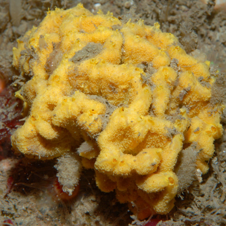
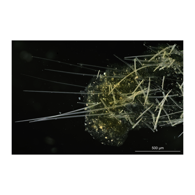
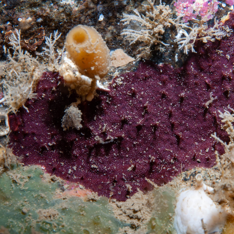
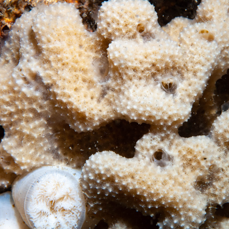
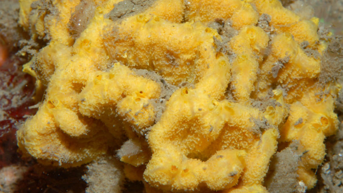
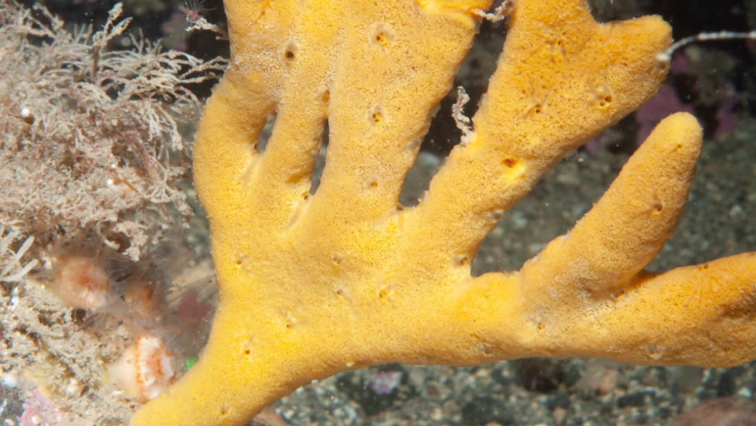
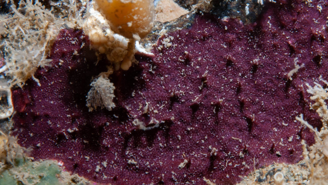
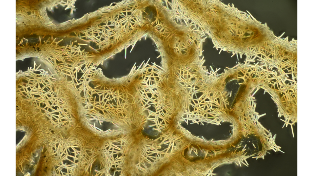
Sponges are tricky to identify as you need to use the formation of the sponge spicule, which makes up their skeleton, to identify the species correctly. These spicules typically range in size from 1mm - .01 mm, and therefore need to be viewed with a microscope which can multiply x400 times. The actual sponge can be quite large, usually 5-50 cm, and often brightly coloured. Yellow, orange and red are the most common colours of sponges.
The Marine Conservation Society guide was developed to help identify the species of sponges in underwater photographs, based on their skeletons and spicules. For each sponge that was added to the guide, a specimen was collected and added to a museum collection. Most specimens are now kept at the Ulster Museum or the Natural History Museum in London.
Since 2005, the Ulster Museum's sponge collection has been greatly expanded by a series of collecting expeditions.
In 2005, the Rathlin Sponge project carried out 6 weeks of fieldwork around Rathlin Island. Rathlin was chosen because the sponges had already been sampled during the Ulster Museum's Northern Ireland Sublittoral Survey in the 1980s and in dives in intervening years, and a number of specimens were thought to represent species which had never been named.
Rathlin has very rich sponge communities because it has strong tidal streams and steep rocky habitats. This combination makes for ideal habitats for many marine invertebrates by bringing abundant planktonic food to animals which are attached to the seabed.
In 2006-7, more sponges were collected from around Northern Ireland as part of an investigation into species of conservation concern. Collection sites included the Maidens, which have some similarity of habitats to Rathlin, and Strangford Lough, with more sheltered habitats. In 2008-10, a similar project to collect sponges using the same methodology of diving, underwater photography, and preservation as museum specimens, was carried out at sites in western Scotland, Wales and the Channel Islands. This expanded the collection to 6000 specimens, with most of these accompanied by underwater photographs.
For these projects, only a small part of the sponge was collected. As sponges have no internal organs, a piece of sponge from the edge of an individual organism gives the full set of spicules needed for identification, allowing the sponge to grow back where it has been sampled.
In 2022, we started a project to digitise our sponge collection. This involved photographing the microscope slides which were made for the identification of each specimen.
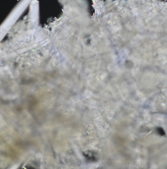
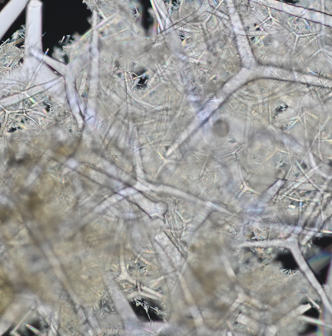
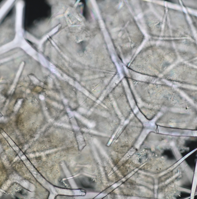
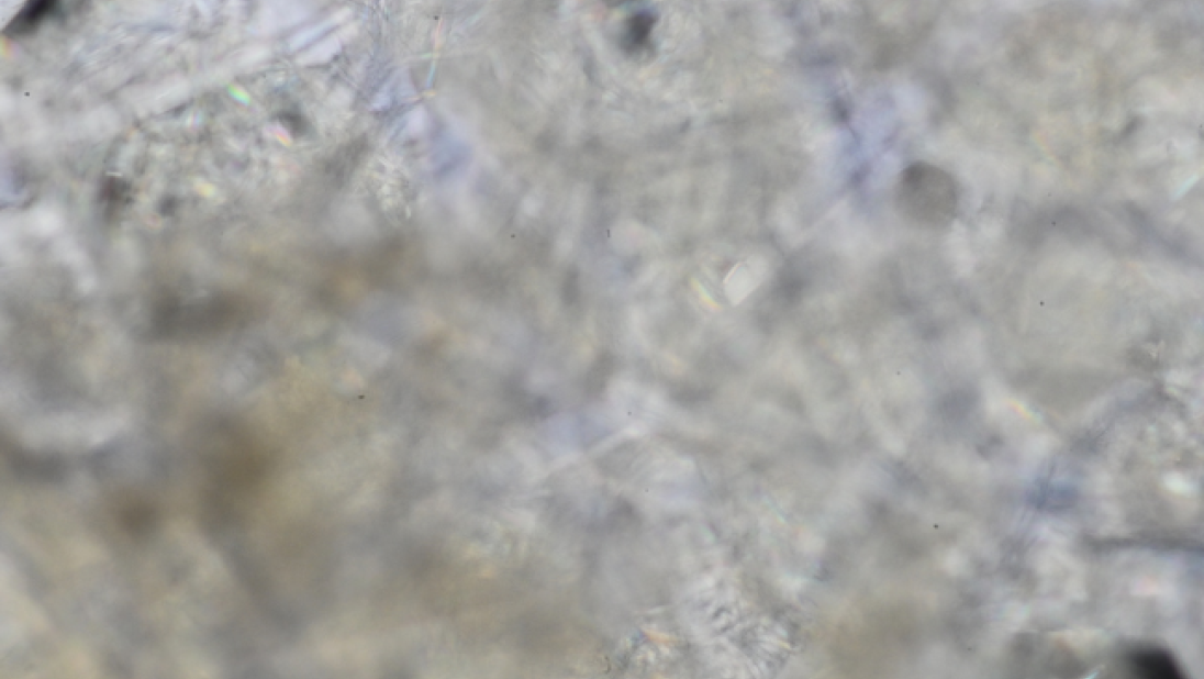
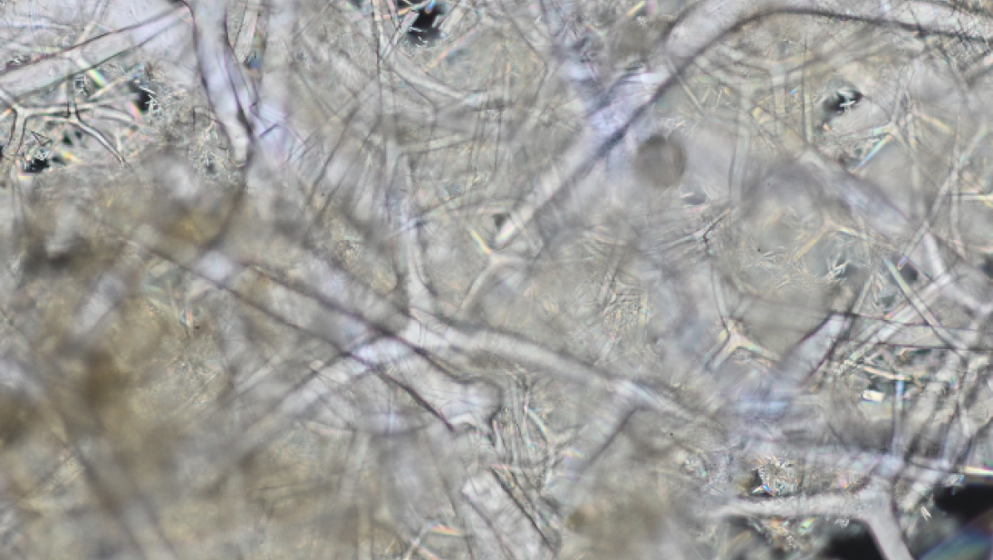
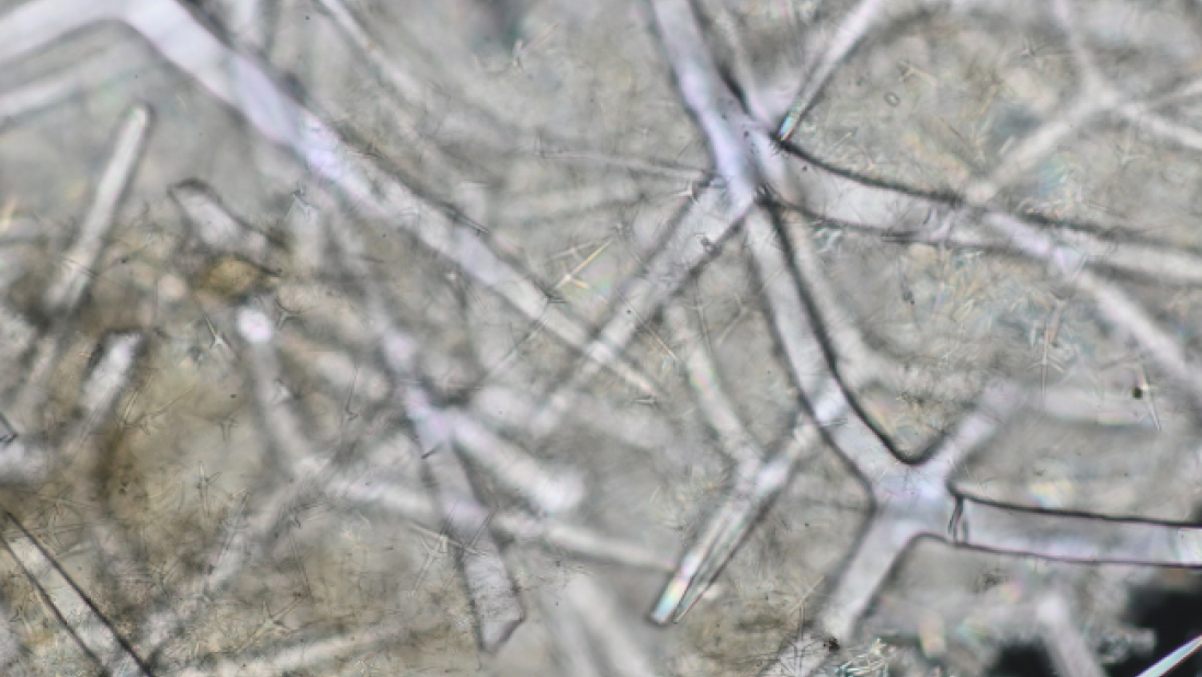
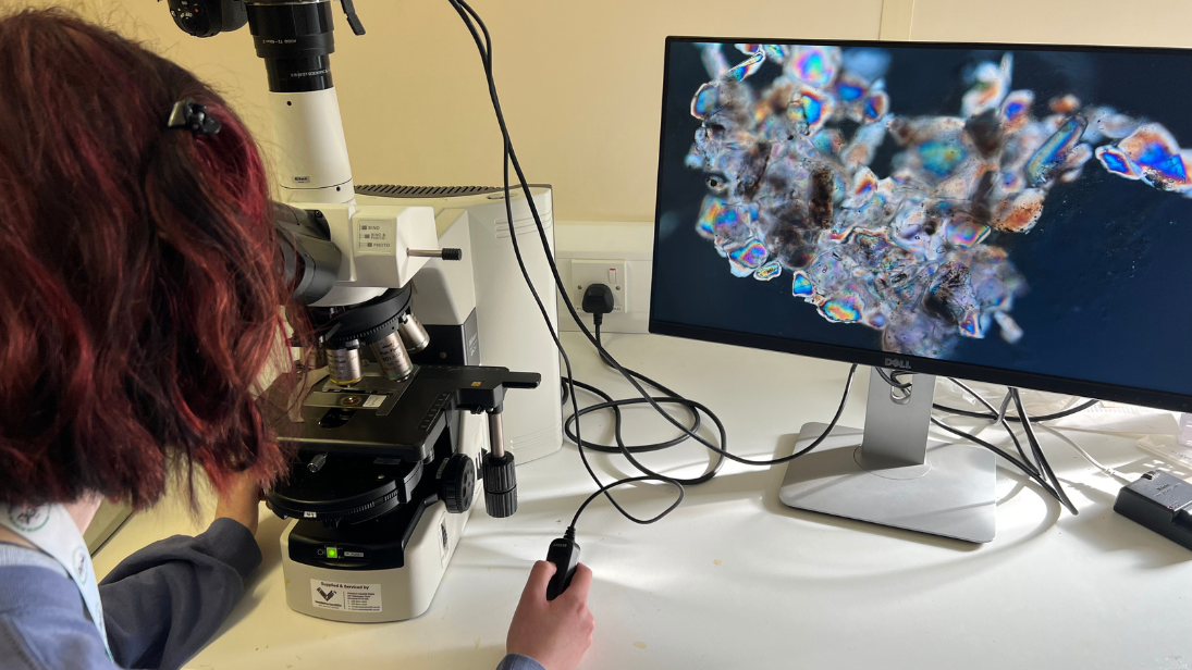
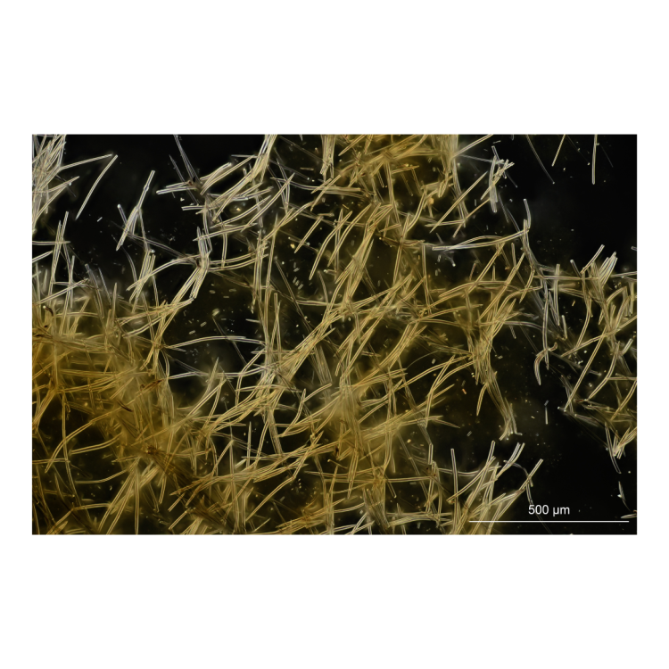
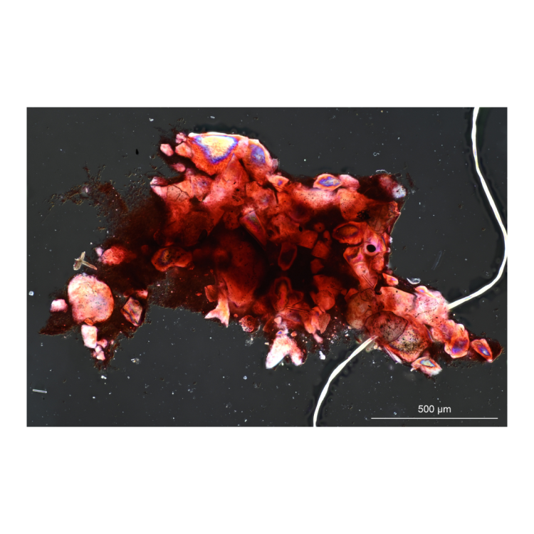
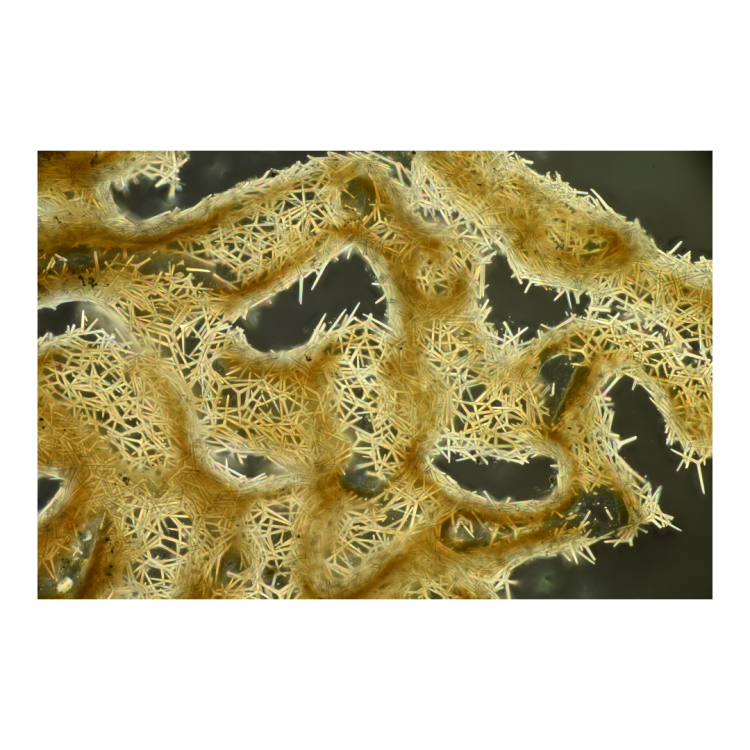
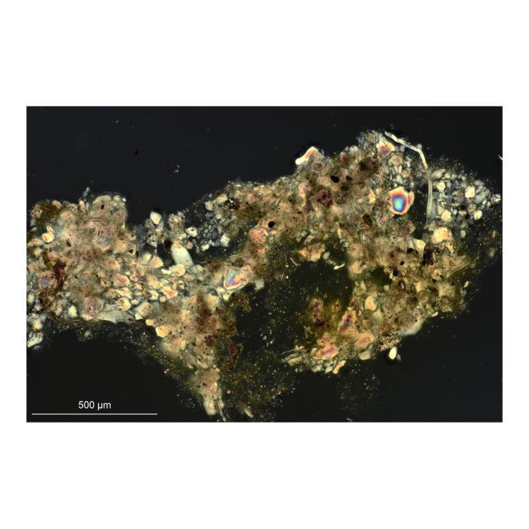
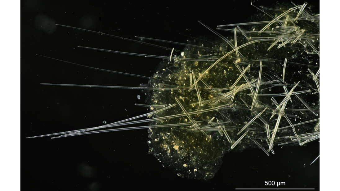
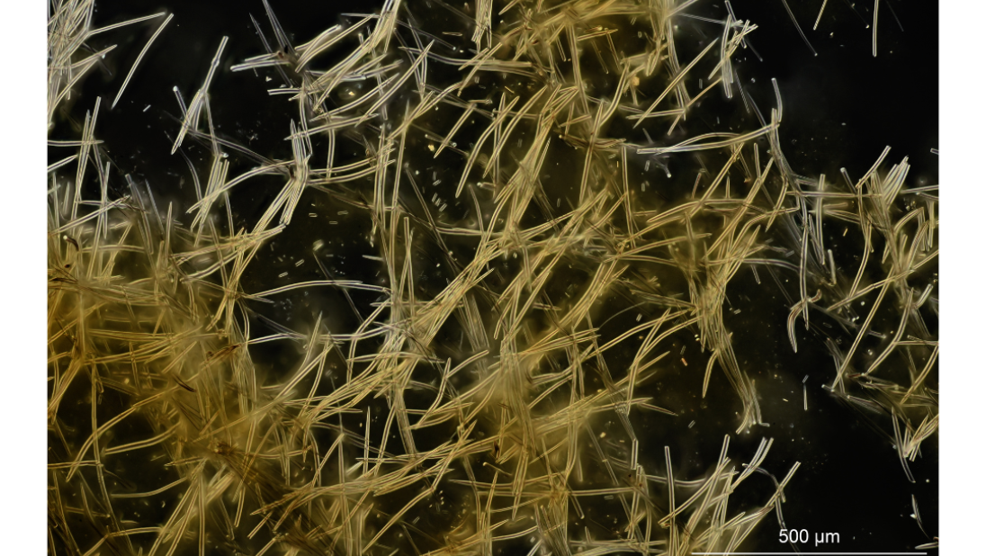
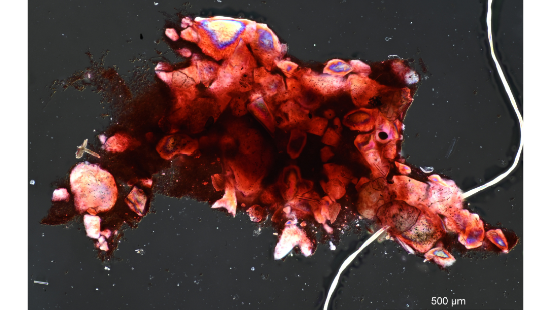
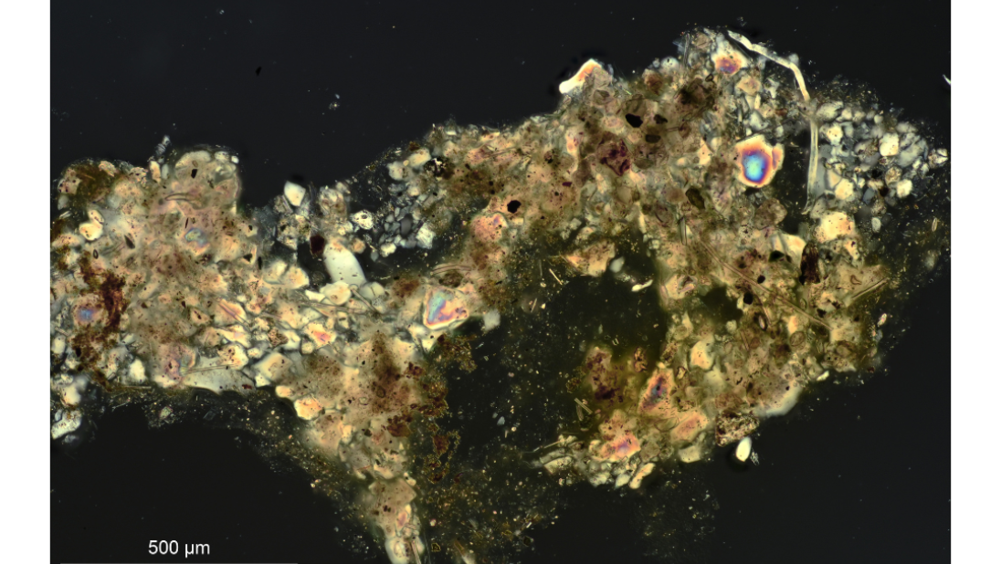
Sponge spicules are made of silica or calcium carbonate, making them transparent. Special lighting techniques are needed to see them clearly down a microscope. For this project, we used a microscope fitted with differential interference contrast optics to take the photographs. It uses two polarising filters set at right angles to cut out the light background, and the spicules then appear bright against a black background. The microscope also has very shallow depth of focus, so only part of a spicule is in focus at a time.
To create images with depth and sharp focus, a series of images are gathered as the microscope is focused slowly through the specimen.
These images are then combined using Helicon focus to stack the images, creating a composite with in-focus parts of each image preserved. The end product is a set of photographs of each species of sponge that illustrate its spicules and the way the spicules are arranged in the skeleton.

These images are added to the existing website and will make it easier for people who want to identify sponges to compare with their own samples. Currently, the website has drawings of the spicules for most species; Scanning Electron Microscopy of some types of spicules; and descriptions. These can be hard to interpret for beginners in sponge identification, who will be looking at a microscope slide with a light microscope and will not easily see the skeleton arrangement or the size differences in different types of spicule.
The images we take are CC-BY copyright so that they can be used on sites such as iNaturalist and the World Porifera database, as well as hosted on our collections website.
Join Dr Helen James and her team on a field trip in Ballinderry, to collect freshwater insects and research species diversity.
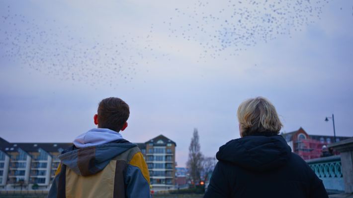
Conor McKinney, Founder and Chair of Wild Belfast, re-tells the beautiful and poignant story of the murmurations disappearance, and return.
Learn about the snow butterflies in the collection of the Ulster Museum!